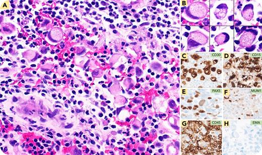
A 41-year-old man presented with a rapidly progressing firm mass in the right supraclavicular area. A complete blood count was normal apart from leukocytosis and neutrophilia. A positron emission tomography/computed tomography scan showed para-aortic lymphadenopathy and a 5-cm supraclavicular mass, with a biopsy showing large, atypical cells in small clusters with background lymphocytes, histiocytes, granulocytes and plasma cells (panel A, hematoxylin and eosin stain [H&E], 20× objective). These cells exhibited abundant cytoplasm, with vacuolization, and eccentrically displaced nuclei resembling signet ring cells. Representative signet ring cells from different areas of the lesion (panel B, H&E, 40× objective). Rare Hodgkin lymphoma–like cells were also noted. Immunohistochemistry showed that these atypical cells were positive for CD30 (panel C, 40× objective), CD15 (panel D, 40× objective), PAX5 (weak) (panel E, 40× objective), MUM1 (panel F, 40× objective), fascin, and BCL6. They were negative for CD45 (panel G, 40× objective), CD20, CD79a, BOB1, OCT2, CD3, CD43, ALK, EBER, mucicarmine, pancytokeratin, and epithelial membrane antigen (EMA) (panel H, 40× objective). The patient was diagnosed with classic Hodgkin lymphoma (CHL).
Signet ring morphology in neoplastic lymphocytes has been reported in various non-Hodgkin lymphomas (eg, follicular lymphomas, diffuse large B-cell lymphomas). To our knowledge, this unusual morphology has not been previously described in the literature in CHL. This case adds CHL to the list of lymphomas rarely presenting with this morphology. Clinicopathological and immunohistochemical evaluation is needed to establish the diagnosis.
For additional images, visit the ASH Image Bank, a reference and teaching tool that is continually updated with new atlas and case study images. For more information, visit https://imagebank.hematology.org.


This feature is available to Subscribers Only
Sign In or Create an Account Close Modal