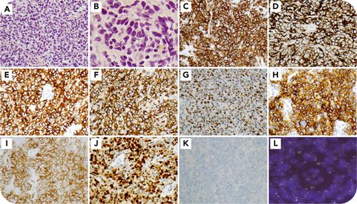
A 73-year-old woman, previously diagnosed with peripheral T-cell lymphoma (PTCL), presented with groin lymphadenopathy. Biopsy showed effaced architecture with pleomorphic intermediate to large lymphoid cells (panel A, 40× objective; panel B, 100× objective, hematoxylin and eosin [H&E] stain). Immunophenotyping revealed diffuse expression of CD2, CD3 (panel C, 40× objective), CD4, CD5, CD20 (panel D, 40× objective), CD30 (panel E, 40× objective), CD15 (panel F, 40× objective), BCL6 (panel G, 40× objective), ICOS (CD278) (panel H, 40× objective), PD-1 (CD279) (panel I, 40× objective), IRF4/MUM1 (panel J, 40× objective) and was negative for CD7, CD8, CD10, ALK-1 (panel K, 40× objective), T-cell cytotoxic markers, CD79a, PAX5, and EBER. Fluorescence in situ hybridization confirmed IRF4/DUSP22 gain in 37 of 200 nuclei (panel L, ZytoLight SPEC IRF4, DUSP22 Dual Color Break Apart Probe, ≥3 fusions), leading to a diagnosis of PTCL not otherwise specified (NOS) with DUSP22alt.
This case uniquely demonstrates coexpression of T follicular helper (TFH) markers, CD30, CD15, and CD20, an uncommon immunophenotype in PTCL-NOS, posing diagnostic challenges and potential misclassification as other T-cell or B-cell lymphomas. As DUSP22 inhibits T-cell activation, its gain may explain aberrant PD-1 and ICOS expression, biomarkers associated with T-cell activation pathways. The presence of TFH markers in DUSP22alt PTCL expands its known immunophenotypic spectrum, emphasizing the importance of comprehensive immunophenotyping and cytogenetics for accurate diagnosis.
For additional images, visit the ASH Image Bank, a reference and teaching tool that is continually updated with new atlas and case study images. For more information, visit https://imagebank.hematology.org.


